resources
How to gather references for medical illustration
By Nancy Ji • Published on Mar 2, 2022
Reference gathering is a fundamental skill for medical illustrators.
Not only does this help us understand the topic better, but it also helps us get better visual results. It can be a time-consuming process, but it is essential to get it right. In today’s article, we want to show you some of the resources we use in our research process.
Medical references
Primary source
Primary sources are first-hand accounts of a topic created by those who were directly connected to the subject matter. For medical illustrators, some of the primary sources we use can be:
- Photos/Videos of the surgeries or device
- Operation notes
- Academic research articles
- Medical imaging (CT, MRI, Xrays)
- Pathology photos (gross anatomy or histology)
- Technical drawings
Secondary source
Secondary sources are materials on a topic created by those who were not directly involved in the actual event. For medical illustrators, some of the secondary sources we use can be:
- Textbooks
- Review articles
- Edited works
- Anatomy apps
- Animations
- Medical models
PSA: For a full list of resources we recommend, check out our free eBook here:
Download Our eBook
Resources to guide your next project
In a previous blog post, we recommended some books on human anatomy that are helpful to our work. Unlike photos, medical illustrations offer more clarity for the viewers by emphasizing information selectively.
However, as medical illustrators, we still try our best to stay faithful to the science we communicate. Primary sources motivate us to observe the original subject matter and look past what the illustrator has designed. This process allows us to come up with unique visual solutions.
Visual analogy
Once we finish gathering medical references, we also need to collect visual references. These help us represent our subject matter more realistically.
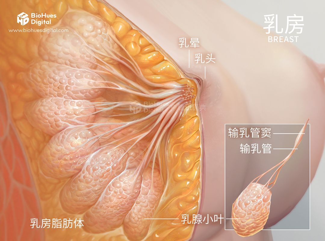
When we need to paint a specific texture or understand the 3D form of a microscopic structure, we try to find organic objects to look at other than the human body. These images are much easier to come by (and sometimes we buy them from the grocery store to study from!) We call them visual analogies. The search can take a little creativity, but the results can be fascinating:
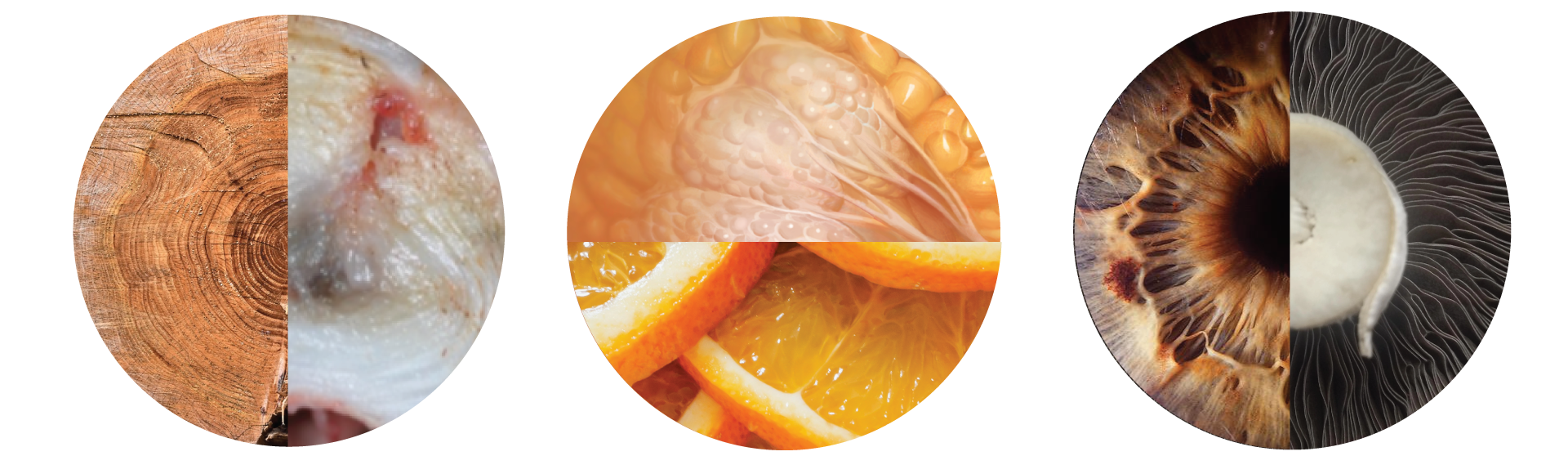
Left: Tree rings vs. annulus from an intervertebral disc. Middle: Breast tissue vs. orange pulp. Right: Iris of an eye vs. the bottom of a mushroom cap.
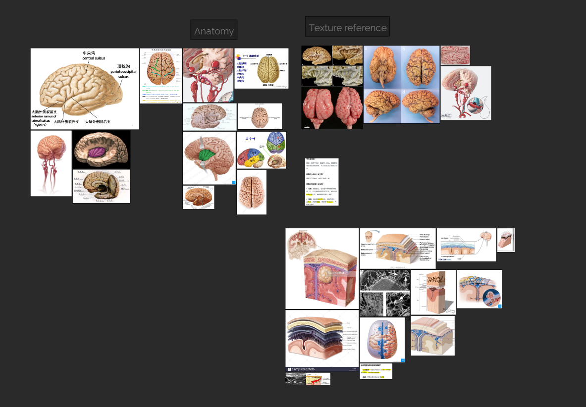
Organizing references
At BioHues, we use PureRef across our team, and it has become one of the tools we cannot live without. Pureref allows us to collect visual references, jot down notes, and save everything in a compact file. (My favourite feature is its “Always On Top” option. This floating window saves me a ton of screen space on my single-monitor setup.)
A huge Thank You to Natascha and Viktor at Idyllicpixel, who created this beautiful application!
Conclusion
At BioHues, we have training sessions to help our team develop good research habits. Over time, we have collected many useful resources. You can download a list of these resources below:
Download Our eBook
Resources to guide your next project
Click on the image to see the digital anatomy atlas we created

About the Author
Nancy Ji is a medical illustrator and the co-founder of BioHues Digital. She holds a Master’s Degree from the Biomedical Communications program at the University of Toronto. Nancy’s specialty is medical animation as well as project management. She wants to advance health equity through accessible medical visuals.